
Leave Your Message
Biological Membrane Infection describes microbial invasion of living barriers, including skin, mucous membranes, and tissue linings. These surfaces are not passive walls. They contain immune cells, protective lipids, mucus, and resident microorganisms. When this balance changes, bacteria, fungi, or viruses may attach, multiply, and damage nearby cells. The result can range from a small inflamed wound to a serious infection affecting deeper tissue.
J. William Costerton, a pioneer in biofilm research, stated, “Biofilms are the dominant mode of bacterial growth in nature.” This observation helps explain why some infections persist. Microbes can form slimy communities on moist surfaces, dental tissue, chronic wounds, or medical devices. The matrix may reduce antibiotic penetration and shield organisms from immune responses. Small details matter. A warm wound. Thick mucus. A poorly cleaned catheter.
Types of Biological Membrane Infection may be classified by location, organism, or infection pattern. Common categories include cutaneous membrane infections, respiratory membrane infections, gastrointestinal membrane infections, and reproductive or urinary membrane infections. Some remain localized, while others spread through damaged barriers. Viral infections may enter cells directly, whereas bacterial and fungal infections often depend on adhesion and tissue disruption.
This classification is useful, but it is not perfect. Several infections involve more than one membrane or change category over time. Accurate diagnosis therefore requires clinical examination, laboratory testing, and attention to symptoms. Redness, swelling, discharge, pain, fever, or a persistent sore should not be interpreted alone. Understanding the infection type supports safer treatment decisions and more reliable prevention.
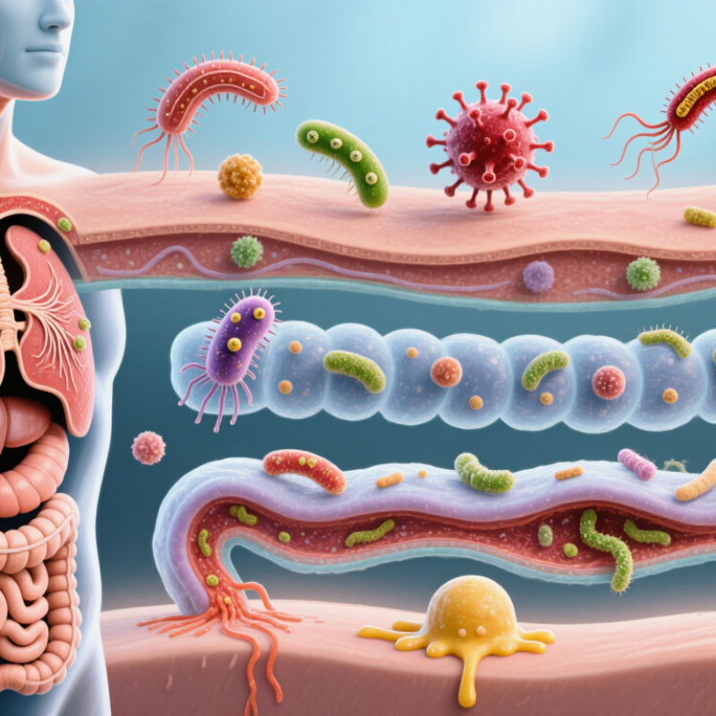
What Is Biological Membrane Infection and Its Types?
Biological membrane infection describes microbial invasion, colonization, or inflammation involving a body membrane. The term is broad, not one single diagnosis. It may involve mucous membranes, skin barriers, serous linings, or specialized tissue surfaces. Clinicians usually define the infection by location, organism, and tissue response.
Common examples include infections of the oral, nasal, intestinal, urinary, and reproductive mucosa. The conjunctiva can also be affected. Serous membrane infections, such as inflammation around the lungs or heart, may become medically serious. Bacteria, viruses, fungi, and parasites can cause different patterns. Some organisms attach to surfaces and form biofilms, which can resist immune clearance and complicate treatment.
Terminology matters. Colonization means microbes are present without clear tissue damage. Infection suggests invasion, multiplication, or a harmful host response. Inflammation may occur with or without infection. These concepts are often confused, even in general health writing. Diagnosis may involve physical examination, microscopy, cultures, molecular tests, and imaging. Test selection depends on the membrane involved and the suspected cause. Symptoms can include redness, discharge, pain, swelling, odor, or impaired function. Yet symptoms alone cannot identify the organism reliably. A careful assessment should consider recent antibiotics, immune status, hygiene, trauma, and exposure history. The phrase “membrane infection” remains useful, but its boundaries are imperfect and require clinical context.
| Dimension | Core Information | Typical Examples | Clinical or Biological Relevance |
|---|---|---|---|
| Working definition | An infection involving a biological membrane, such as the skin, a mucosal lining, a serous membrane, or the membrane of an individual cell. | Skin infection, conjunctival infection, respiratory mucosal infection, or infection of a cell membrane by an intracellular pathogen. | The term is descriptive rather than a single standardized disease category. The affected membrane and the causative organism determine the diagnosis. |
| Biological membrane | A selectively permeable lipid-and-protein structure that separates cells, tissues, or body compartments. | Plasma membrane, epithelial barrier, mucosal lining, pleura, peritoneum, and meninges. | Membrane damage can disrupt barrier function, fluid balance, signaling, and local immune defense. |
| Primary site: skin | Infection of the epidermis, dermis, hair follicles, sweat glands, or associated soft tissue. | Impetigo, folliculitis, cellulitis, dermatophytosis, and viral vesicular eruptions. | Common findings include redness, warmth, swelling, pain, itching, scaling, pustules, or vesicles. |
| Primary site: mucosa | Infection of moist epithelial membranes lining the respiratory, gastrointestinal, genitourinary, or ocular surfaces. | Conjunctivitis, oral candidiasis, infectious rhinitis, pharyngitis, and cervicitis. | Mucus, ciliary movement, epithelial integrity, and local antimicrobial factors influence susceptibility and disease progression. |
| Primary site: serous membrane | Infection involving membranes that line closed body cavities and cover internal organs. | Pleuritis associated with infection, peritonitis, and inflammation of the pericardial membrane. | These infections may cause fluid accumulation, severe inflammation, impaired organ movement, or systemic illness. |
| Bacterial membrane infection | An infection caused by bacteria that colonize, invade, or damage a membrane or its underlying tissue. | Bacterial cellulitis, bacterial conjunctivitis, impetigo, and bacterial peritonitis. | Bacteria may produce toxins, enzymes, capsules, or biofilms that support adherence and immune evasion. |
| Viral membrane infection | A viral infection affecting epithelial or cellular membranes, often through receptor-mediated entry and intracellular replication. | Herpetic skin lesions, viral conjunctivitis, influenza-like respiratory infection, and viral gastroenteritis. | Viruses can alter membrane proteins, cause cell fusion, disrupt membrane integrity, or trigger programmed cell death. |
| Fungal membrane infection | An infection caused by yeasts or filamentous fungi that affects keratinized tissues or mucosal surfaces. | Dermatophytosis, oral candidiasis, vulvovaginal candidiasis, and fungal nail infection. | Fungal growth is influenced by moisture, temperature, skin barrier disruption, immune status, and antimicrobial exposure. |
| Parasitic membrane infection | An infection in which parasites or parasitic organisms inhabit, penetrate, or damage epithelial and mucosal surfaces. | Scabies affecting the skin and protozoal infections affecting intestinal or genitourinary mucosa. | Symptoms may result from direct tissue injury, immune reactions, nutrient competition, or secondary infection. |
| Local infection | An infection confined mainly to one membrane or nearby tissue. | Localized impetigo, uncomplicated fungal skin infection, or isolated conjunctivitis. | Local symptoms are usually dominant, although progression to deeper tissue or systemic disease remains possible. |
| Invasive infection | An infection that crosses the membrane barrier and extends into deeper tissue, body cavities, blood, or other organs. | Necrotizing soft-tissue infection, invasive fungal disease, and peritonitis with systemic spread. | Invasive disease can cause sepsis, organ dysfunction, tissue necrosis, or life-threatening inflammatory responses. |
| Biofilm-associated infection | A microbial community attached to a surface and enclosed in a self-produced extracellular matrix. | Chronic wound infection, dental plaque-associated disease, and infections involving implanted medical materials. | Biofilms may reduce antimicrobial penetration and impair immune clearance, contributing to persistence or recurrence. |
| Barrier disruption | Loss or weakening of membrane protection caused by wounds, burns, inflammation, dryness, maceration, or underlying disease. | Infected cuts, burn wound infection, eczema-associated infection, and postoperative wound infection. | Barrier disruption increases microbial entry, fluid loss, inflammation, and the risk of secondary infection. |
| Common signs and symptoms | Inflammatory or tissue-specific changes occurring at the infected membrane. | Redness, warmth, pain, swelling, itching, discharge, ulceration, scaling, blisters, odor, or fever. | Symptoms vary by site and pathogen; severe pain, rapidly spreading redness, confusion, or breathing difficulty requires urgent medical assessment. |
| Diagnostic approach | Clinical examination supported by laboratory testing when the diagnosis is uncertain, severe, recurrent, or systemic. | Microscopy, culture, nucleic-acid testing, antigen testing, blood tests, imaging, or tissue sampling. | Testing should be selected according to the suspected pathogen, anatomical site, disease severity, and treatment history. |
| Prevention principles | Measures that preserve membrane integrity and reduce exposure to infectious organisms. | Hand hygiene, wound care, respiratory etiquette, safe sexual practices, appropriate vaccination, and avoiding unnecessary antimicrobial use. | Prevention is most effective when combined with management of chronic conditions and prompt care of damaged tissue. |
Note: “Biological membrane infection” is a broad descriptive expression rather than one universally defined medical diagnosis. The specific membrane, infectious organism, depth of involvement, and clinical presentation should guide evaluation and treatment.




 Phone
Phone Send Email
 whatsapp